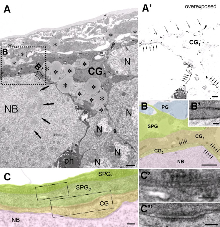
Fig 4. Ultrastructural features of the LD accumulating superficial cortical glial cells.
(A) HRP-labeled superficial cortical glial cell (CG1), encapsulating a mitotic neuroblast (NB, arrows: chromosomes) and other neurons (N). Note the abundance of LDs. (A’) Same picture as shown in panel A but overexposed during acquisition to reveal the DAB stained processes (arrows) at low magnification. Note that a phagosome (ph, labeled in panel A) accumulates HRP. (B, B’) Higher magnification pseudocolored image taken from the dashed areas in panel A. DAB stained glial membranes are indicated (arrows). Two overlapping cortex glial processes (CG1 and CG2) insulate a mitotic neuroblast (NB). (C) Organization of the glial layers encapsulating neuroblasts. (C’) Subperineurial glial cells (SPG1-2) are connected to each other through septate junctions. (C”) A subperineurial (SPG2) and a superficial cortex glia (CG) are connected with an adherens junction. Scalebar: (A, A’, B) 1 μm, (B’,C, C’, C”) 100nm.

