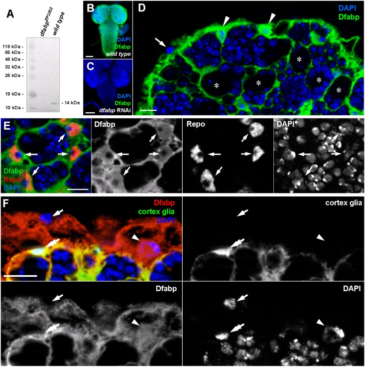
Fig 5. Light microscopic localisation of the Drosophila fatty acid binding protein (Dfabp) in third instar larvae.
(A) Western blot performed on total protein extracts from dfabp 3252 homozygous mutant and wild type larvae. The Dfabp antibody labels a single band at 14kDa in the wild type sample, while no labeling is observed in samples from mutants. Immunohistochemistry on wild type (B), and on dfabp RNAi (C) third instar larval brains using the Dfabp antibody (green). Nuclei are stained with DAPI (blue). Note the absence of staining in RNAi silenced compared to wild type animals. (D) Higher magnification image of the dorsomedial part of the central brain. The Dfabp antibody reveals a thin network, between neuroblasts (asterisks) and their daughter cells. Two Dfabp-positive (arrowheads), and one unlabeled soma (arrow) are visible at the brain surface. (E) Double labeling against Dfabp (green) and the glial-specific protein Repo (red). Note that Dfabp is present in the cytosol and in the nucleus of glial cells (arrows). (F) Double labeling for cortex glia-GFP (green) and Dfabp (red). Perineurial cells (arrow) are double negative for Dfabp and GFP. Subperineurial cells (arrowhead) are positive for Dfabp and negative for GFP. Cortex glial cells (double arrow) are double positive for Dfabp and GFP. Scalebar: B, C:100 μm; D, E, F, F’: 10 μm.

