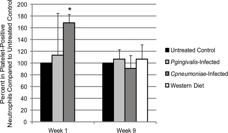
Fig 6. Heterotypic aggregate formation in ApoE-/- mice with infection and diet.
Whole blood samples taken on Week 1 (24 h after the last P. gingivalis challenge and 4 days after exposure to C. pneumoniae; n = 3 for each group) and on Week 9 (n = 3 for each group) and dual stained for platelet marker (CD41) and neutrophil marker (Ly6G). The percent of platelet-positive neutrophils were determined through flow cytometry and normalized to Untreated Control at each timepoint. Data is normally distributed and analyzed using an ANOVA. *p<0.05 compared to Untreated Control at Week 1.

