
Fig 7. The protein expression of Rev1 was upregulated in response to DNA damage.
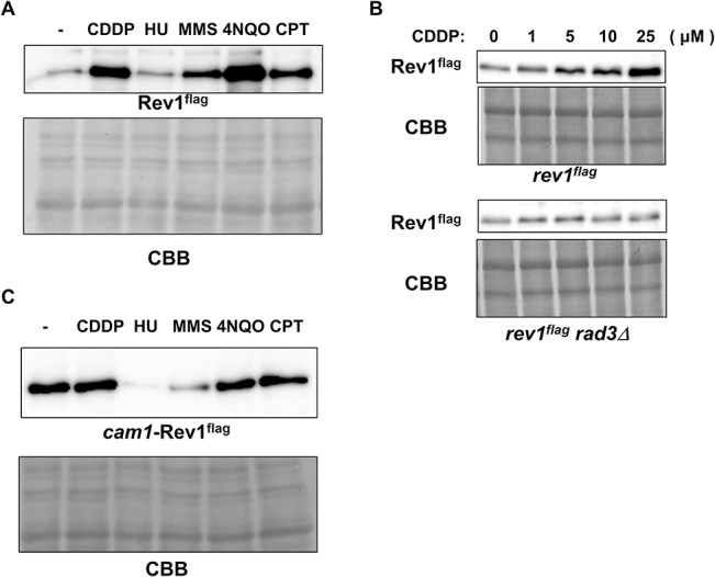
A, Rev1 protein expression after mutagen treatment. At the logarithmic growth phase, wt cells harboring flag-tagged rev1 were treated with no drug (-), 50 μM cisplatin (CDDP), 10 mM hydroxyurea (HU), 0.008% MMS, 500 nM 4NQO, or 40 μM camptothecin (CPT). After a 4-h incubation, cells were harvested. Whole cell extracts were prepared by the boiling method, and protein levels were examined by western blotting. The upper panel represents flag-tagged Rev1, and the lower panel shows CBB staining of the membrane. B, The upregulation of Rev1 was dependent on Rad3. At the logarithmic growth phase, rev1flag and rev1flag rad3Δ strains were treated with 0, 1, 5, 10, or 25 μM cisplatin for 3 h. Cells were then harvested, and whole cell extracts were prepared by the boiling method. Extracts were then subjected to western blotting. The upper panels show Rev1 and CBB staining of the rev1flag strain, and the lower panels show those of the rev1flag rad3Δ strain. C, The promoter region was important for the upregulation of Rev1 after DNA damage. At the logarithmic growth phase, cam1-rev1 cells were treated with no drug (-), 50 μM cisplatin (CDDP), 10 mM hydroxyurea (HU), 0.008% MMS, 500 nM 4NQO, or 40 μM camptothecin (CPT). After a 4-h incubation, cells were harvested, and whole cell extracts were prepared by the boiling method. Protein expression was then examined by western blotting. The upper panel shows Rev1, and the lower panel shows CBB staining.