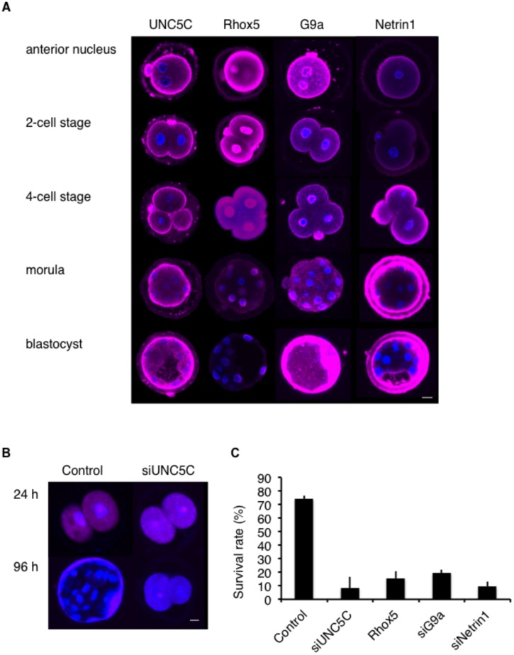
Fig 6. G9a decreases Rhox5 expression.
A. Representative images showing UNC5C, Rhox5, G9a, and Netrin-1 (each shown by pink fluorescence) in the anterior nucleus, 2-cell, 4-cell, morula, and blastocyst stages. Bar: 10 μm. B. Representative images showing mCherry (pink fluorescence) in murine embryos at 24 or 96 h after electroporation with mCherry only (Control) or with mCherry and siUNC5C (siUNC5C). Bar: 10 μm. C. Survival rates of murine embryos at 96 h after electroporation with mCherry only (Control), mCherry and siUNC5C (siUNC5C), mCherry and Rhox5 (Rhox5), mCherry and siG9a (siG9a) or mCherry and siNetrin-1 (siNetrin-1).

