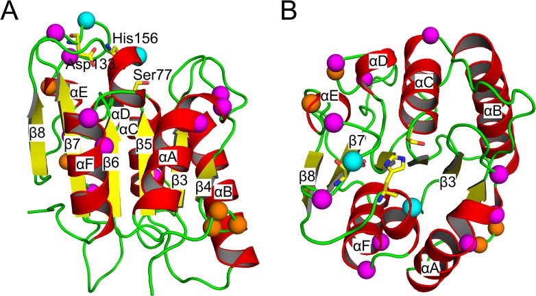
Fig 2. Cartoon representation of wild type BsLipA with mutated residues indicated by spheres of their Cα atoms (mutations from Rao et al. [44–48]: magenta; Reetz et al. [42, 49]: orange; mutations common in both data sets: cyan).
The catalytic triad (Ser77-Asp133-His156) is shown in stick representation with yellow carbons. The protein is colored according to secondary structure (α-helices: red; β-sheets: yellow; loops: green). The right view (B) differs from the left (A) by an anti-clockwise rotation of ~90° about a horizontal axis. All figures of BsLipA structures were generated with PyMOL (http://www.pymol.org).

