
Figure 1.
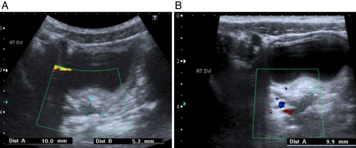
Ultrasound scan and colour Doppler of the pelvis showing enlarged and vascular seminal vesicles, the right (A) being more enlarged than the left (B).
Official websites use .gov
A
.gov website belongs to an official
government organization in the United States.
Secure .gov websites use HTTPS
A lock (
) or https:// means you've safely
connected to the .gov website. Share sensitive
information only on official, secure websites.
Ultrasound scan and colour Doppler of the pelvis showing enlarged and vascular seminal vesicles, the right (A) being more enlarged than the left (B).