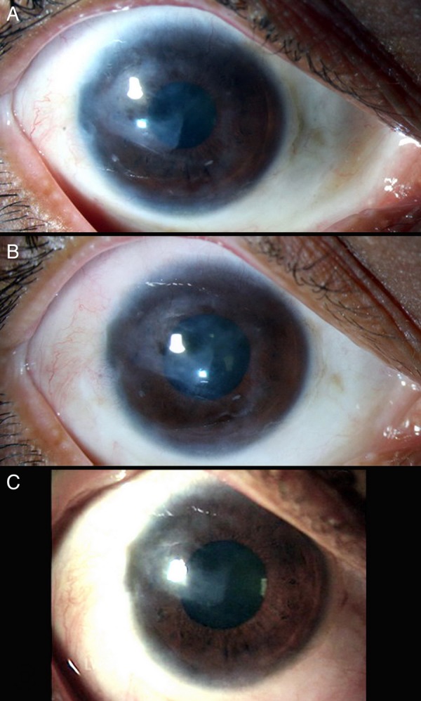
Figure 3.

Serial slit lamp photographs of the right eye at (A) 5 months, (B) 10 months and (C) 27 months (last follow-up) postoperatively, showing gradual dissolution of limbal explants, reduction in peripheral corneal vascularisation and increased clarity of cornea with maintenance of a healthy epithelium.
