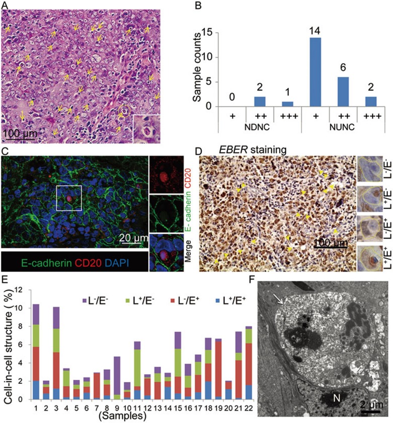
Figure 1.
Cell-in-cell structures formed between B lymphocytes and nasopharyngeal ECs in NPC tissues. (A) Typical heterotypic cell-in-cell structures in one NPC tissue sample. Heterotypic cell-in-cell structures were indicated by yellow arrows. (B) Frequency of heterotypic cell-in-cell structures in NDNC (n = 3) and type 2b (III) NUNC (n = 22) determined by hematoxylin-eosin staining. The cell-in-cell frequency was scored with four scales: “−”, 0%; “+”, 1%-5%; “++”, 5%-10%; “+++”, 10%-15%. (C) Representative images of lymphocyte-nasopharyngeal EC cell-in-cell structures in a human NPC sample with co-staining of E-cadherin (green) and CD20 (red). DAPI staining (blue) indicated the nucleus. (D) EBER staining of NPC samples. Four types of heterotypic cell-in-cell structures were presented in the right lane. L−/E−: EBER− lymphocytes/EBER− ECs; L+/E−: EBER+ lymphocytes/EBER− ECs; L−/E+: EBER− lymphocytes/EBER+ ECs; L+/E+: EBER+ lymphocytes/EBER+ ECs. (E) Statistics of four types of heterotypic cell-in-cell structures in individual specimen indicated by different colors. (F) A representative TEM image of heterotypic cell-in-cell structure in tissue sections of NPC. In a typical cell-in-cell structure, the internalized B cell (indicated by a white arrow) was surrounded by the vacuole and the deformed nucleus (N) of the EC.

