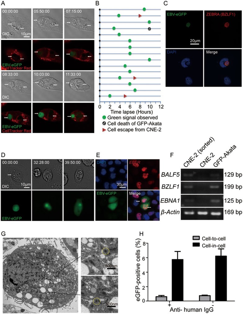
Figure 2.
EBV-carrying GFP-Akaka cells transmit virus to CNE-2 cells through cell-in-cell interaction. (A) Time tracking observation of EBV activation in GFP-Akata cells within CNE-2 cells. CNE-2 cells (pre-stained with CellTracker Red dye) were co-cultured with GFP-Akata cells at a ratio of 1:10 and observed using LSM 710 confocal microscope. It was notable that internalized GFP-Akata cells became green (GFP-positive), which indicated EBV activation, at time 07:15:00. Time was indicated as hour:minute:second. Scale bar,10 μm. (B) The fate of individual GFP-Akata cell was indicated, including the appearance of GFP (green circle), undergoing cell-in-cell death (black circle) or escaping from CNE-2 cells (red triangle). Data analysis was performed for 12 h with an 1 h interval. (C) Expression of ZEBRA (red) in an EBV-activated GFP-Akata cell (green) inside a CNE-2 cell (indicated by DAPI) as determined by immunofluorescence staining. (D) Time tracking analysis of GFP diffusion into CNE-2 cells. The CNE-2 cells were co-cultured with GFP-Akata cells at a ratio of 1:5 and observed using DMI6000B fluorescence microscope. The target CNE-2 cells became GFP positive at time 39:50:00, which was the indicator of EBV infection. Time was indicated as hour:minute:second. Scale bar, 10 μm. (E) Distribution of EBER (red) and GFP (green) in cell-in-cell structures. After incubation of GFP-Akata cells with CNE-2 cells for 12 h, the free GFP-Akata cells were removed by washing with PBS. The sorted CNE-2 cells with cell-in-cell structures were hybridized with the probe to EBER for ISH. (F) PCR analysis of the indicated viral mRNAs in the sorted CNE-2 cells with cell-in-cell structures. GFP-Akata cells served as positive control while non-treated CNE-2 cells served as negative control. (G) TEM observation of EBV virions (yellow circle) in in-cell infected CNE-2 cells. The in-cell infected CNE-2 cells were obtained by FACS sorting after co-culturing with GFP-Akata cells at a ratio of 1: 10 and maintained with G418-containing culture medium for 2 weeks. (H) A summary of cell-in-cell- and cell-to-cell-mediated EBV transmission. CNE-2 cells were co-cultured with GFP-Akata cells (with or without anti-human IgG treatment) for 24 h. The frequencies of GFP-positive CNE-2 cells freely or with cell-in-cell structures were analyzed by fluorescence microscopy.

