Figure 5.
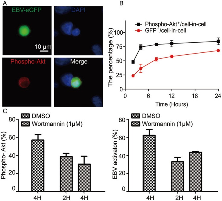
Autonomous activation of EBV inside CNE-2 cells depends on the PI3K/AKT signaling pathway. (A) GFP-Akata cells were co-cultured with CNE-2 cells for 12 h and the phosphorylation of AKT inside GFP-Akata cells was detected by immunofluorescence staining using anti-phospho-AKT antibody followed by DAPI staining. The images were captured under confocal laser scanning microscope. (B) Proportional kinetics of phospho-AKT+ GFP-Akata and GFP+ CNE-2 cells within cell-in-cell structures. GFP-Akata cells were co-cultured with CNE-2 cells. The percentages of phospho-AKT+ GFP-Akata and GFP+CNE-2 cells were analyzed by confocal laser scanning microscopy at the indicated times. The experiments were performed three times independently. (C) The PI3K signaling inhibitor Wortmannin was added to the culture medium after cell-in-cell structure formation (2 or 4 h after co-culture of GFP-Akata and CNE-2 cells). The percentages of phospho-AKT+ GFP-Akata cells (left) and GFP+ GFP-Akata cells (right) within cell-in-cell structures were determined by fluorescence microscopy 24 h after the treatment.

