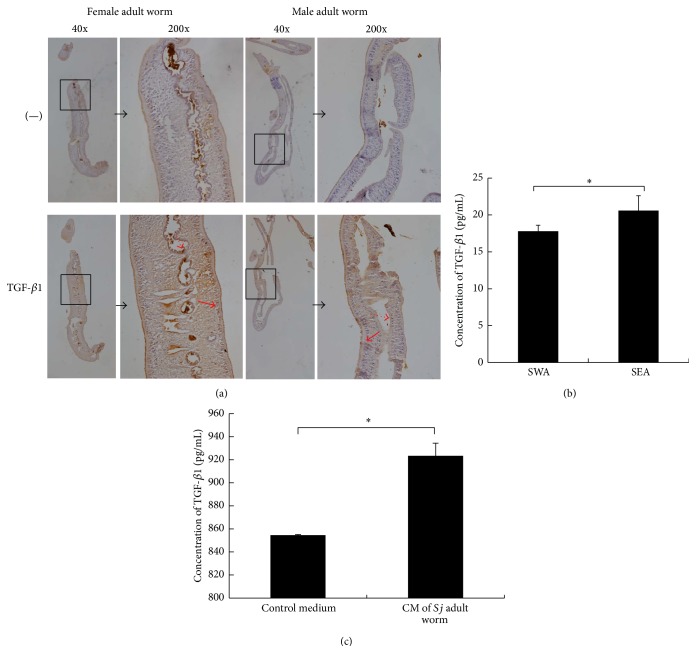
Figure 3.
TGF-β1 protein was expressed in Sj and was secreted. (a) Sj adult female and male worms were fixed in paraformaldehyde, embedded in paraffin, and then sliced and stained for TGF-β1. Representative staining (brown) is shown at 40x magnification (large panel) and ×200 (inset). Dotted red arrow: gut epithelial cells; solid red arrow: subtegumental cells. (b) Equal amounts of Sj soluble adult worm antigen and soluble egg antigen were tested for TGF-β1 by ELISA. Data were shown as means ± SD. Experiment was performed four times (∗ P < 0.05; and ∗∗ P < 0.01 compared with negative PBS control). (c) Twenty pairs of adult worms were freshly collected, washed thrice using PBS, transferred into 2 mL sterile RPMI 1640 medium supplemented with 1 mM glutamine, 1000 units/mL penicillin, and 1000 μg/mL streptomycin for 2 h, and finally cultured in 2 mL sterile RPMI 1640 medium supplemented with 20% FBS, 1 mM glutamine, 100 units/mL penicillin, and 100 μg/mL streptomycin for 16 h. Sj worm culture medium was collected for TGF-β1 detection by ELISA, and condition medium (sterile RPMI 1640 medium supplemented with 20% FBS, 1 mM glutamine, 100 units/mL penicillin, and 100 μg/mL streptomycin) was used as control. Experiment was performed four times (∗ P < 0.05; and ∗∗ P < 0.01 compared with control medium without Sj adult worms).

