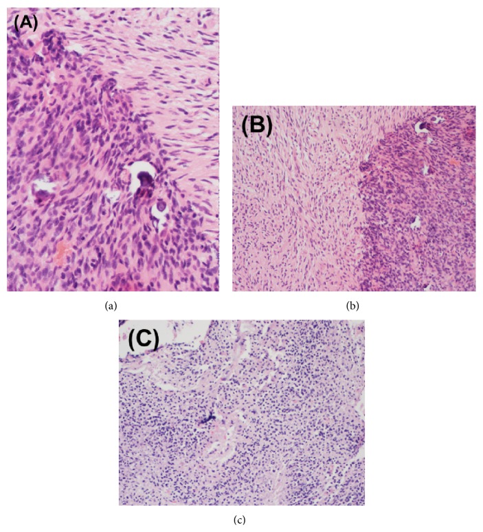
Figure 1.
Surgical pathological specimens from (a) initial craniotomy and (b-c) repeat craniotomy for recurrent intracranial disease. Histopathological analysis of the initial (a) right temporal lobe specimen demonstrated glioblastoma with small cell features. The pathological specimen after second resection (b) demonstrated active glioblastoma, almost entirely viable with <5% necrosis. Following the third and final (c) craniotomy for locally recurrent disease, predominantly viable (approximately 15% necrosis) active glioblastoma was identified. Molecular studies were negative for MGMT methylation and EGFR amplification was detected by FISH.

