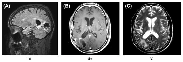
Figure 3.

Intracranial imaging during final course of salvage therapy with bevacizumab and carboplatin. (a) FLAIR, (b) T1-weighted axial, and (c) T2-weighted axial magnetic resonance imaging demonstrating stable intracranial disease at time of respiratory decline. Postsurgical hyperintensity surrounds area of resection in the right temporal and occipital lobes communicating with the right occipital horn, unchanged with respect to prior imaging.
