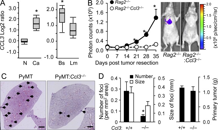
Figure 3.
Loss of CCL3 in stromal cells suppresses pulmonary metastasis of human and mouse breast cancer cells. (A) Oncomine search results for CCL3 are shown. (left) CCL3 transcript abundance in stroma of breast cancer (Ca) and normal tissue (N). P = 2.61 × 10⁻¹⁶; fold change, 3.078. (right) CCL3 levels in basal-like (Bs) and luminal-like (Lm) invasive breast carcinoma showing increase in basal cancers. P = 2.28 × 10⁻⁴; fold change, 1.747. (B) Lung metastatic burden from orthotopic mammary tumors was quantified (n = 3/genotype, 3 independent experiments). The tumors were developed by MDA231:4175TGL human breast cancer cells in nude mice transplanted with Rag2−/− or Ccl3−/−Rag2−/− BM cells. Plots show normalized photon flux in the lung over time. Data are means ± SEM. *, P < 0.05. Representative images of mice at 35 wk are shown (right). (C) Representative H&E stained microscopy images of the lung from PyMT (n = 8) and PyMT:Ccl3−/− (n = 7) mice with spontaneous metastatic foci (Arrowheads) are shown. Bars, 1 mm. (D) Number and size of lung metastatic foci (left) and primary tumor weights (right) were assessed in autochthonous PyMT (n = 8) or PyMT:Ccl3−/− mice (n = 7) at 27–30 wk of age (4 independent experiments). Data are means ± SEM. *, P < 0.01.

