Figure 5.
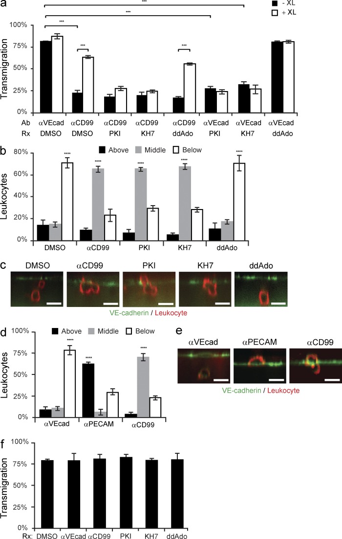
Inhibiting sAC or PKA blocks leukocyte transmigration. (a) TEM assays were performed using HUVECs pretreated with either anti–VE-cadherin or anti-CD99, as well as DMSO, PKI, KH7, or ddAdo. Before fixation, either GαM IgG (cross-linking antibody, XL) or GαRb IgG (control) was added to samples for 10 min (b and c) PBMCs were added to HUVECs pretreated with DMSO, anti-CD99, PKI, KH7, or ddAdo. Samples were stained with anti–VE-cadherin (EC) and anti-CD18 (leukocyte). Confocal images were taken to assess the site of blockade. Leukocytes were scored as being above the endothelium, blocked partway through, or migrated below HUVEC monolayers. (d and e) HUVECs were pretreated with anti–VE-cadherin, anti-PECAM, or anti-CD99. PBMCs were allowed to transmigrate for 1 h. Cells were then fixed, stained, imaged, and analyzed (as described above). (f) Eluate control TEM assays were performed as previously described (Mamdouh et al., 2009). In brief, HUVECs were pretreated with anti–VE-cadherin, anti-CD99, PKI, KH7, ddAdo, or DMSO for the duration of the incubation in blocking experiments. Cells were then washed and fresh media was added to samples. HUVECs were incubated at 37°C for 1 h (the duration of the normal blocking experiments). The media was collected from each well. The eluate media contains all of the inhibitor that would have eluted out of the cultures over the duration of the blocking experiment. PBMCs were resuspended in the eluate media, added to untreated HUVECs, and incubated at 37°C for 1 h. Cells were subsequently fixed and analyzed. Bars, 10 µm. Images are representative of two (e) or three (c) independent experiments. Numerical values are the average of two (d and f) or three (a and b) independent experiments. Error bars represent SEM (***, P < 0.001; ****, P < 0.0001; Student’s t test [a] and ANOVA [b, d, and f]).

