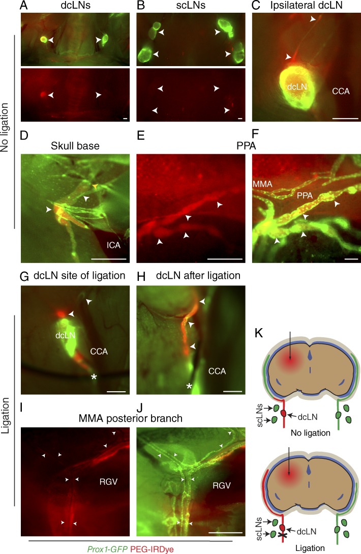
Figure 2.
Dura mater lymphatic vessels drain brain ISF into dcLNs. (A–J) Analysis of lymphatic outflow routes of cerebral ISF by fluorescent stereomicroscopy in Prox1-GFP (green) mice 1 h after PEG-IRDye (red) injection into the brain parenchyma without (A–F) and with (G–J) ligation of the efferent lymphatic vessel of the dcLN. See K for schematic illustration of the experimental setup and summary of the results with and without ligation. (A and B) dcLNs and scLNs (both indicated with arrowheads) showing preferential filling of the ipsilateral dcLN but no filling in the scLNs. (C) Drainage into the ipsilateral dcLN via the efferent carotid lymphatic vessels (arrowheads). CCA, common carotid artery. (D) Internal carotid artery (ICA) and adjacent lymphatic vessels (white arrowheads) immediately below the osseous skull, showing drainage from the skull (yellow arrowhead). (E and F) Lymphatic vessels around the pterygopalatine artery (PPA), showing tracer uptake by the dura mater lymphatic vessels (arrowheads) only in the basal parts of the skull, nearby their exit site. MMA, middle meningeal artery. (G) Placement of a suture around the efferent lymphatic vessel (asterisk) of the dcLN. Arrowheads, afferent lymphatic vessels. (H) Afferent lymphatic vessel of the dcLN after ligation (asterisk), showing bulging of the afferent vessels (arrowheads). (I and J) Lymphatic vessels around the posterior branch of the MMA, showing increased filling of lymphatic vessels after ligation, extending above the retroglenoid vein (RGV) level. n = 2–3/group. Data are representative of two independent experiments. Bars: (A–E and G–J) 500 µm; (F) 100 µm.

