Figure 3.
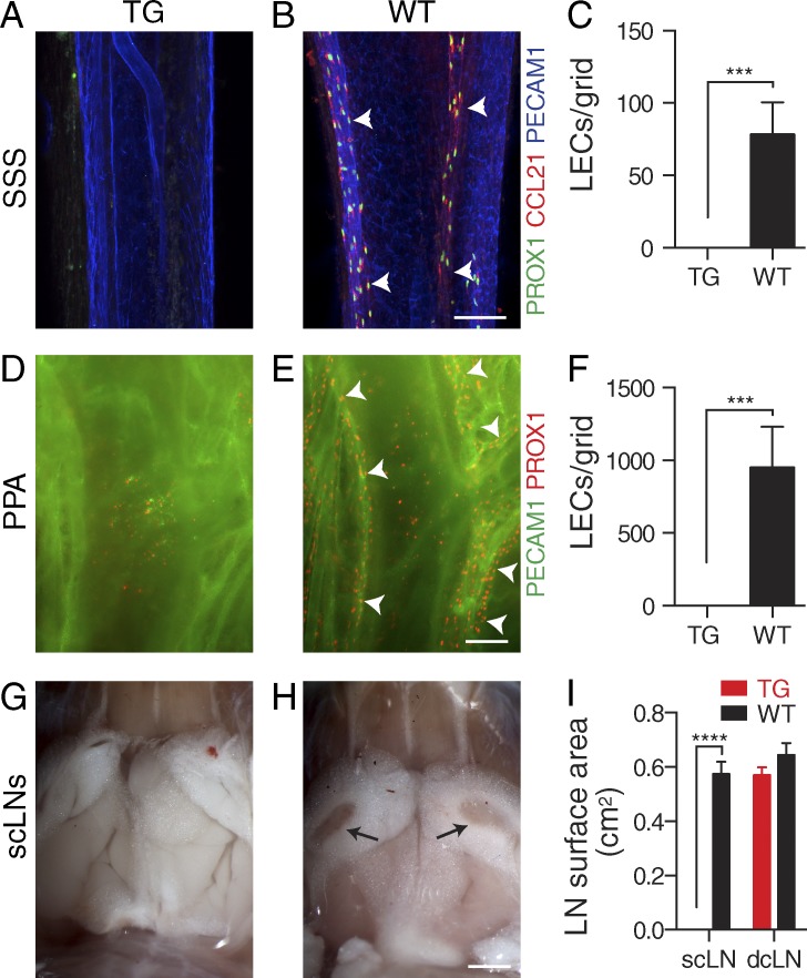
Absence of dural lymphatic vasculature in K14-VEGFR3-Ig TG mice. (A–F) Analysis of dura mater lymphatic vasculature in K14-VEGFR3-Ig TG and WT littermate control mice. (A–C) Immunofluorescence of the superior sagittal lymphatic vessels (arrowheads) for PECAM1, PROX1, and CCL21 (A and B) and quantification of PROX1+/CCL21+ lymphatic ECs (LECs)/grid (C). (D–F) Immunofluorescence of the pterygopalatine and middle meningeal lymphatic vessels (arrowheads) for PECAM1 and PROX1 (D and E) and quantification of PROX1+ LECs/grid (F). (G–I) Stereomicroscopic photographs showing the absence of the scLNs (arrows) in the TG mice (G and H) and quantification of the (mean left/right) scLN and dcLN surface areas (I). Micrographs of the dcLNs are shown in Fig. 4 C. (A–F) n = 3 (TG) and 4 (WT). (G and H) n = 4/group. Data are representative of two independent experiments. Bars: (A, B, D, and E) 100 µm; (G and H) 2 mm. Error bars indicate SD. Statistical analysis: two-tailed Student’s t test (C and F) and two-way ANOVA followed by Šídák’s post-hoc test (I). ***, P < 0.001; ****, P < 0.0001.

