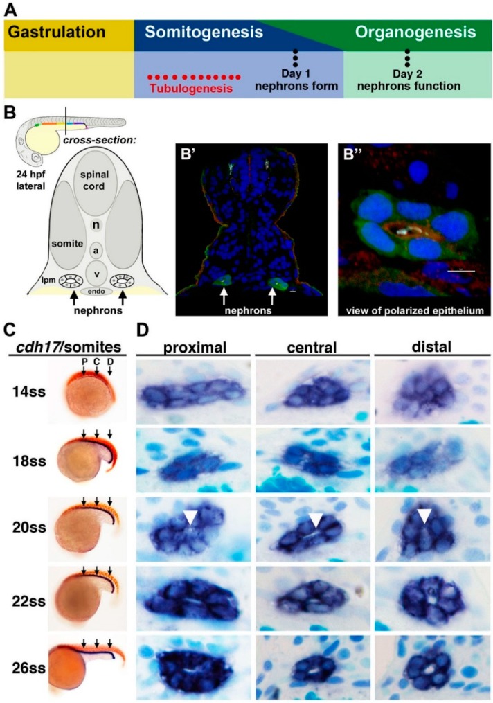
Figure 2.
Tubulogenesis of the zebrafish pronephros. (A) The timing of tubulogenesis is coincident with the stages of somitogenesis and organogenesis of the embryo; (B–B”) At 24 hours post fertilization (hpf), the two nephrons have formed distinct tubule lumens that can be detected by immunofluorescence to detect green fluorescent protein (GFP), acetylated tubulin (light blue), Prkc ι/ξ (red) and nuclei (DAPI, blue); (C, D) The precise onset of tubulogenesis occurs at the 20 ss, indicated by white arrowheads, along the proximal, central, and distal regions of the nephron territory, with progressive enlargement of the luminal space at 22 and 26 ss. Abbreviations: aorta (a), lateral plate mesoderm (lpm) notochord (n), somite stage (ss) cardinal vein (v), [Figure adapted from [43,57], through terms of the Creative Commons License of the Authors].

