Figure 2.
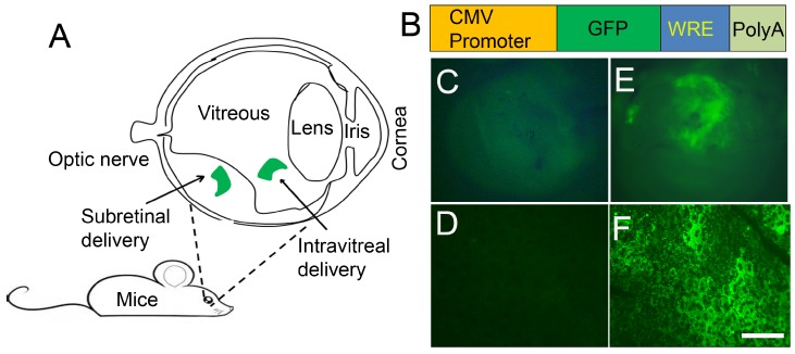
LPD-mediated gene delivery into the retina. Schematic illustration of the eye and route of administration. The most commonly used and preferred mode of administration to retinal layers is subretinal (A). Generation of green fluorescent protein construct under the control of CMV promoter (B). CMV, cytomegalovirus; GFP, green fluorescent protein; WRE, posttranscriptional regulatory element from the woodchuck hepatitis virus; PolyA, polyadenylation sequence; increases the stability of the molecule. Using BalbC mice, we injected the cDNA construct subretinally into one eye. LPD was complexed with CMV-GFP-WRE-PolyA construct. The other eye was injected with LPD, with a control vector without GFP. Seventy-two hours later, eyes were removed and examined for GFP expression under inverted fluorescence microscopy. GFP expression is clearly seen in the GFP-injected eye (E), but not in the control eye (C). Whole RPE flat mounts were prepared and examined for GFP expression under inverted fluorescence microscopy. GFP expression is seen in the GFP-injected eye (F), but not in the control eye (D). Scale bar, 20 µm.

