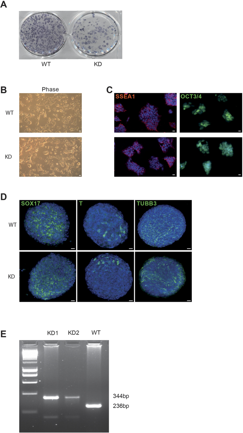
Figure 5. Generation of PKD2 kinase-dead iPS cells.
(A) AP staining of murine embryonic fibroblasts 13 days after infection with a hOSKM encoding lentivirus. KD = PKD2 kinase-dead; WT = Wildtype. (B) Phase contrast microscopy of a feeder-free ESC culture of the respective genotype (C) SSEA1 and Oct 3/4 staining of PKD2 kinase-dead (KD)- and wildtype (WT)-iPSCs. (D) Whole-mount staining of differentiating embryoid bodies (EBs) under non-pluripotency conditions on day 4. EBS are positive for markers of all three germ layers (Endoderm: Sox17; Mesoderm: T; Ectoderm: Tubb3). (E) PCR genotyping of genomic DNA producing products of 236 bp (PKD2-WT) and 344 bp (PKD2 kinase-dead).

