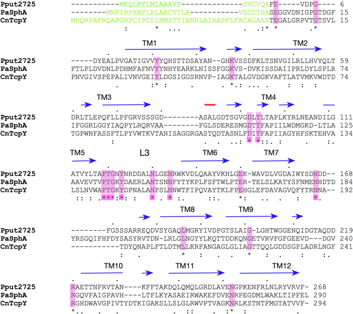
Figure 5. ClustalW sequence alignment of the COG4313 channels Pput2725, C. necator TcpY and P. aeruginosa SphA.
The signal sequences as predicted by SignalP are shown in green. The observed secondary structure elements based on the crystal structure of Pput2725 are indicated (β-strands; blue, helices; red). Identical residues (*) located within the putative lateral exit site are highlighted in pink.

