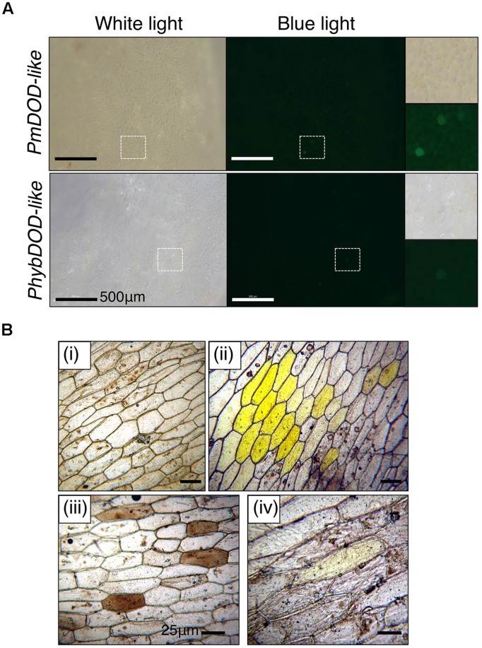
FIGURE 6.
Cell phenotypes in tissue bombarded with DOD-like constructs and fed DOPA. Tissue bombarded with constructs for PmDOD-like and PhybDOD-like. The GFP construct was not included. (A) A. majus petal tissue used. Single green fluorescent cells at low frequency were observed under blue light. A few of these cells were possibly pale pink under white light in the PmDOD-like bombarded sample. Hatched squares show regions depicted at higher magnification in the panels on the right side. Scale bar is the same size in all. (B) White onion scale tissue used. No pigments observed when bombarded with gold particles only (i); conspicuous multicellular yellow zones observed when bombarded with the PmDOD construct (positive control; ii); brown cells (iii) and a rare pale yellow cell (iv) was observed when bombarded with the PhybDOD-like construct. Scale bar = 500 μm (A), 25 μm (B).

