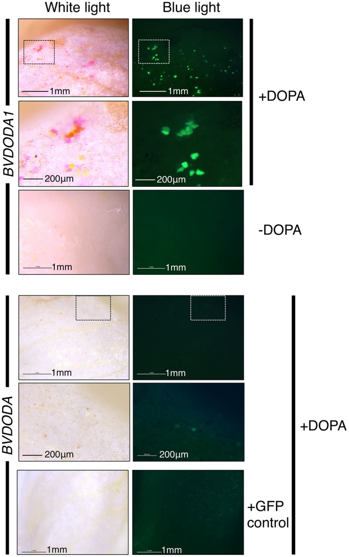
FIGURE 9.
Differential activity of BvDODA and BvDODA1 in a transient expression assay. White petal tissue of A. majus was bombarded with constructs for the genes (driven by the 35S promoter) followed by supplementation with DOPA or water. Representative petal samples are shown. Pigmentation was only visible in BvDODA1 bombarded tissue that was also fed DOPA. The pigmented regions also had fluorescence in blue light characteristic of betaxanthins. The only phenotype observed in BvDODA-bombarded tissue was a small number of cells in the DOPA-fed samples with weak green fluorescence in blue light. Control tissue bombarded with both BvDODA and GFP constructs and fed DOPA lacked pigment formation while GFP fluorescence indicated transformation had occurred. Scale bar = 1 mm. Hashed squares show regions depicted at higher magnification in panels with 200 μm scale bars.

