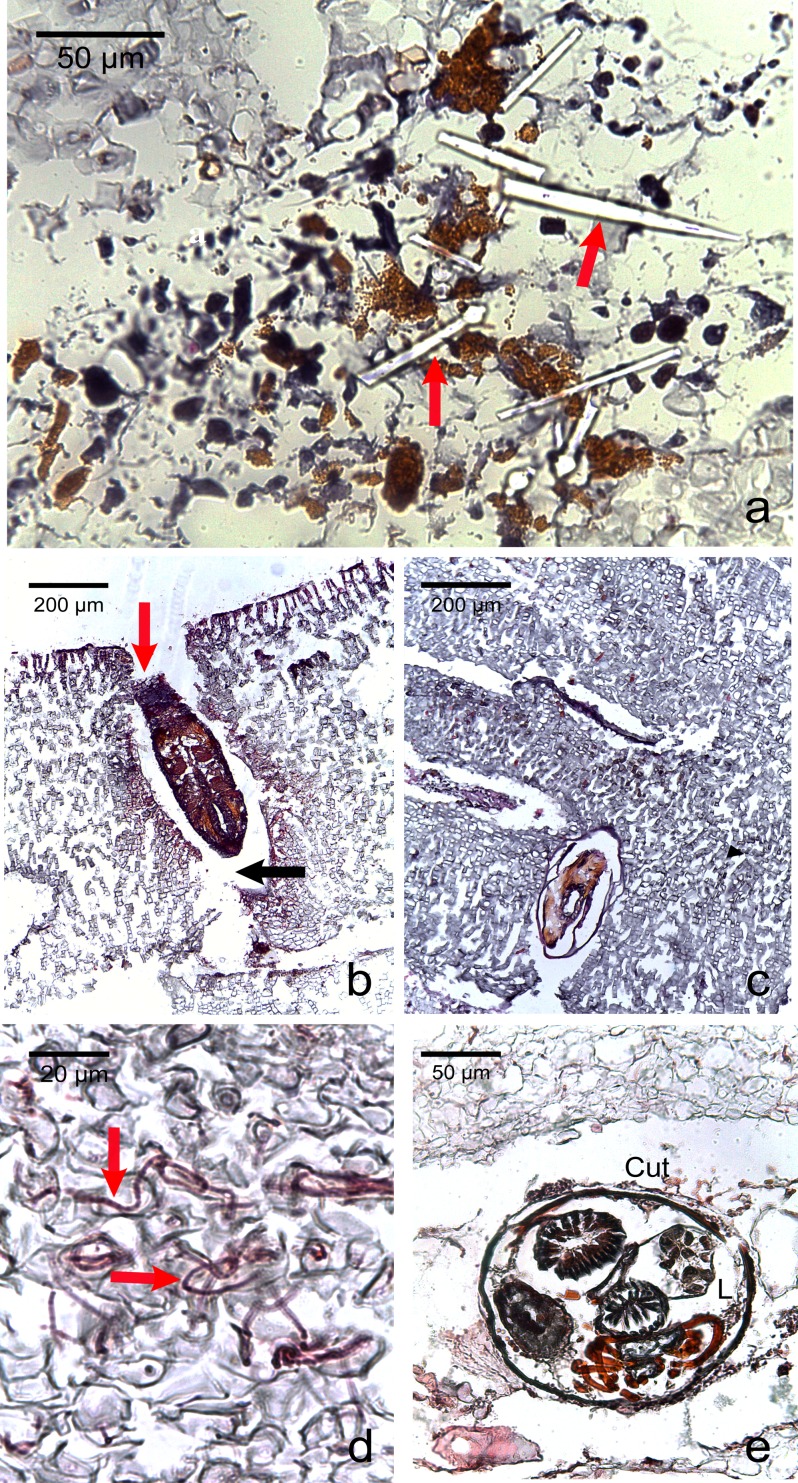
Figure 5. Photomicrographs of the most commonly encountered organisms in healthy and diseased CCA.
(A) Boring sponge characterized by silicaceous spicules (red arrow) (B) Unidentified macroborer. Note the CCA cells lining up the burrow suggesting the growth of the algae around the invader (red arrow) and the acellular space around the organism (black arrow). (C) Unidentified macroborer, possibly a juvenile bivalve. (D) Cyanobacterial trichomes (red arrows); (E) Helminth; Cu, cuticule; L, lumen.

