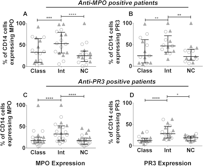
Figure 2. The ANCA autoantigens MPO and PR3 are preferentially expressed on intermediate monocytes.
Peripheral blood was collected from patients with AAV and the percentage of cells expressing cell-surface MPO and PR3 was examined by flow cytometry. The percentage of MPO and PR3 positive cells in each subset is shown for (A–B) anti-MPO+ AAV patients and (C–D) anti-PR3+ AAV patients. Each symbol represents a separate individual. Open circles represent patients in remission and filled triangles patients with active disease. Data are presented as the median and interquartile range. Non-parametric one-way ANOVA (Friedman test) and Dunn’s post-test were used to test for significance (*p < 0.05, **p < 0.01; ****p < 0.0001). Class: classical; Int: intermediate: NC: non-classical.

