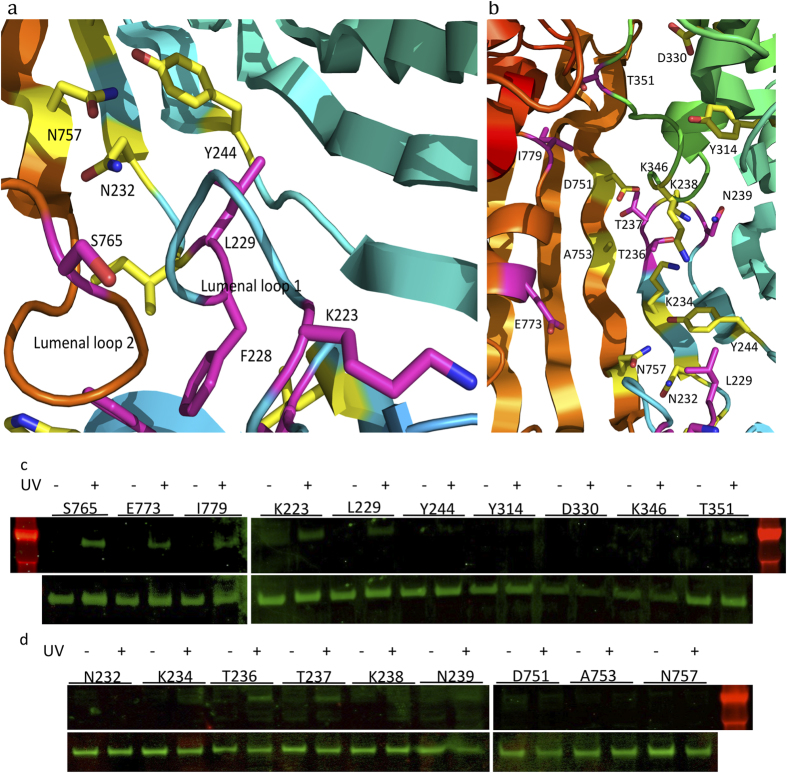
Figure 3. Observation of LptD and LPS complexes at the luminal gate, lumen of the barrel and the extracellular loops.
The positions of residues where cross-linking with LPS was detected are shown in magenta; residue positions with no LPS cross-linking are shown in yellow. a the residues for the incorporation of pBPA at the lumenal gate. b the residues for incorporating pBPA at the lumen of the LptD barrel and the extracellular loops. c and d the top lanes are the detection of the LptD and LPS complexes at the lumenal gate, lumen of the barrel and the extracellular loops, and the bottom lanes are the protein expression levels of the LptD variants. The LptD/LPS complexes were detected at residues K223, L229 and S765 of the lumenal gate, T236, E773, I779 of the lumen of the LptD barrel, T236, N239 and T351 of the extracellular loops 1 and 4.

