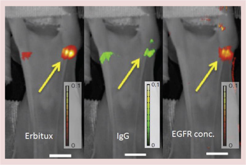
Figure 4. Lymph node images using fluorescent dyes.

Lymph node imaging of tumors with Eribtux labeled with fluorescent reporter IRDye800 and nonspecific reporter IgG labeled with IRDye680, are shown (left and middle) with the processed image of receptor concentration for epidermal growth factor receptor shown (right). Imaging with this approach shows the ability to detect and quantify metastases down to as few as 200 cells in the lymph node [29]. Scale bar: 1 cm.
EGFR: EGF receptor.
