
Fig. 3.
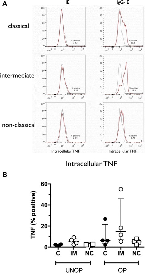
CD14hiCD16+ monocytes produce more TNF compared to other monocytes in response to IE. a Representative histograms showing intracellular TNF staining of monocytes four hours after addition of CS2-IE (IE, left hand panels) or CS2-IE opsonised with rabbit anti-human RBC antibody (IgG-IE, right hand panels). Grey histograms: 4 °C controls, red histograms: 37 °C. b Median (IQR) of intracellular TNF expression in classical (C; solid black circles), intermediate (IM; open circles) and non-classical (NC; open squares) monocytes from four independent experiments using blood from separate donors. IE infected erythrocytes, IQR interquartile range