Fig. 1.
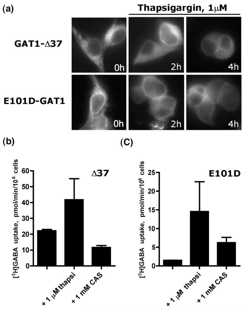
Depletion of calcium from ER or inhibition of ER glucosidases rescues cell surface expression of GAT1-E101D mutant. (a) Fluorescence microscopy of HEK293 cells transfected with GAT1-Δ37 or GAT1-E101D and treated with 1 μM thapsigargin. Cell surface expression of GAT1-E101D is evident after 4 h. (b) Whole-cell [3H]GABA uptake by GAT1-Δ37 or GAT1-E101D (c) following treatment with 1 μM thapsigargin (thapsi) or 1 mM castanospermine (CAS). The data shown are means of two independent experiments; error bars indicate SD.

