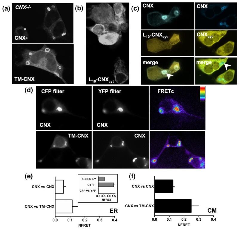
Fig. 3.
The transmembrane portion of calnexin supports targeting to concentric bodies. (a) YFP-tagged calnexin or a truncated form of calnexin lacking the luminal domain (TM-CNX) was visualized using confocal microscopy in transfected cnx−/− MEFs. (b) Confocal images of L10-calnexin-cyt (L10-CNXcyt) construct confirm that it associates with the intracellular membranes. (c) Co-expression of full-length calnexin with the L10-calnexincyt shows that the concentric membranes stained by calnexin contain L10-calnexincyt, but there is no enrichment of the latter (left panel, arrowhead); co-expression of the YFP-tagged soluble C-terminus of calnexin (CNXcyt) with the CFP-tagged calnexin shows that the soluble fragment is excluded from the concentric bodies (right, arrowhead). (d) FRET microscopy and analysis was performed in transfected HEK293 cells as described under Materials and Methods; here, images constructed using only FRETc values are shown. Determination of NFRET was performed in ROIs within (e) the diffuse ER (n=10–15), and (f) multilamellar structures (n=36–89).

