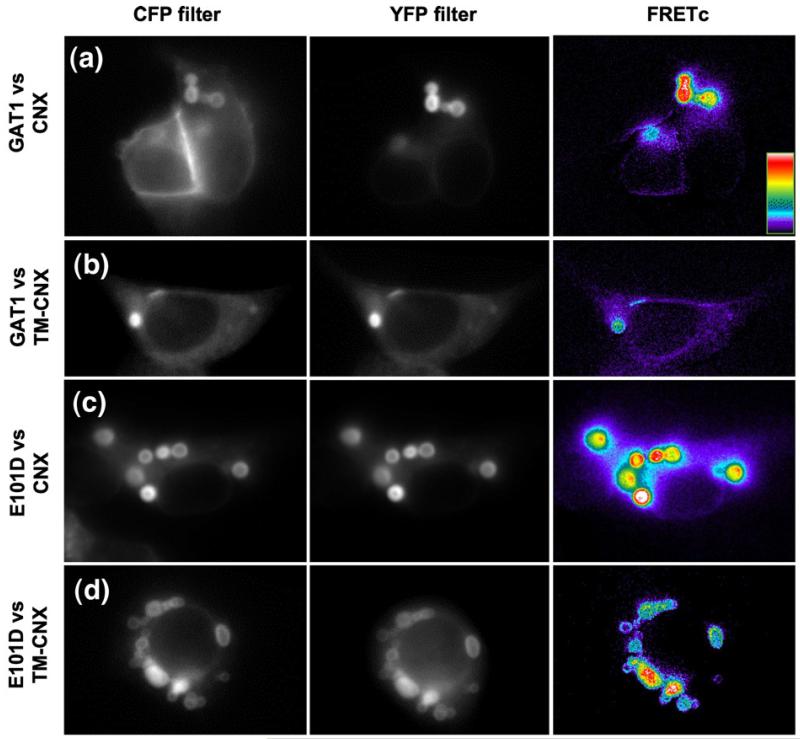
Fig. 6.
GAT1 and E101D colocalize with calnexin and TM-calnexin in concentric bodies. (a–d) HEK293 cells were co-transfected with plasmids driving the expression of CFP-tagged GAT1 (a, b) or of CFP-tagged GAT1-E101D (c, d) and of YFP-tagged calnexin or of YFP-tagged TM-calnexin, as indicated. Proteins were visualized by epifluorescence microscopy as outlined under Materials and Methods. The right-hand panels in each row represent FRETc images (computed as in Fig. 3 according to the equation in Materials and Methods); results for each pair of interacting proteins are summarized in Fig. 8.

