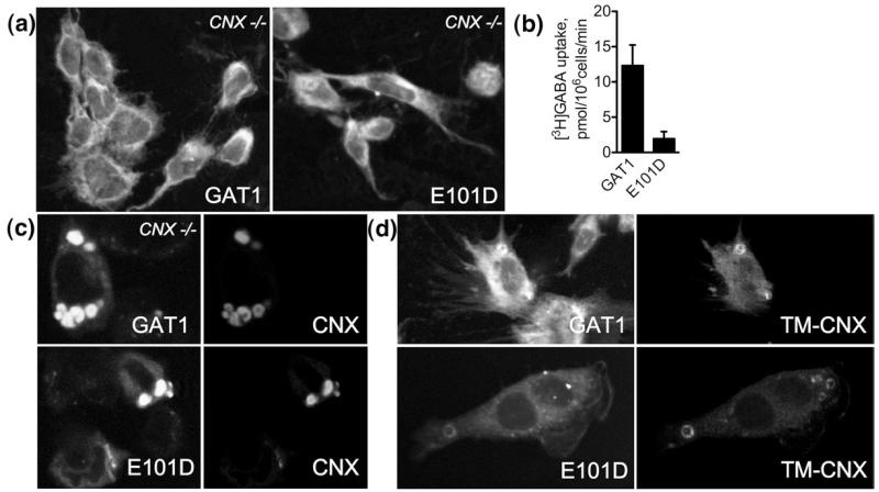
Fig. 7.
GAT1 molecules colocalize with calnexin-derived constructs in concentric membranes in the absence of endogenous calnexin. (a) Confocal microscopy images reveal poor cell surface expression of GAT1 and E101D. cnx−/− MEFs grown on glass coverslips were transfected with the indicated constructs and subjected to confocal microscopy analysis 48 h later. The bulk of the protein is retained inside the cell. (b) Whole-cell [3H]GABA uptake experiments confirm lack of cell surface-expressed GAT1 and E101D. The experiments were performed as described in the legend to Fig. 1 and in Materials and Methods (the bars represent means±SEM from four experiments). (c and d) Confocal images show that GAT1 and E101D are enriched in the concentric membranes upon co-expression of calnexin or TM-calnexin.

