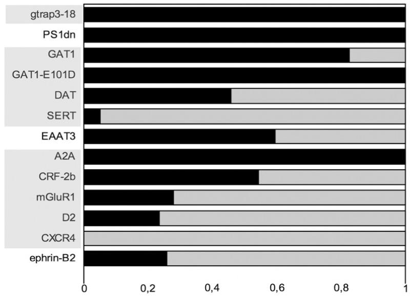
Fig. 9.
Quantification of selective targeting of membrane proteins to concentric bodies. The indicated proteins, tagged with a fluorescent protein, were co-expressed by transient transfection together with either CFP- or YFP-tagged TM-calnexin (as appropriate for dual-wavelength imaging/colocalization experiments) in HEK293 cells. Twenty-four hours later, coverslips with the transfected cells were mounted on an epifluorescence microscope stage and images of CFP and YFP/GFP channel were acquired. Only cells that co-expressed the protein of interest with TM-calnexin construct were analysed. The dark area of the bar indicates the fraction of concentric bodies that were positive for both TM-calnexin and the membrane protein of interest. The light field corresponds to that fraction of concentric bodies containing only TM-calnexin. The abbreviations of the proteins are shaded to indicate separate protein families (NSS, GPCRs, etc.). The n of observed concentric bodies for each co-expressed protein are as follows: GTRAP3-18, 27; PS1-dn, 19; GAT1, 58; GAT1-E101D, 47; DAT, 59; SERT, 58; EAAT3 (excitatory amino acid transporter-3), 37; A2A (A2A adenosine receptor), 66; CRF-2b (corticotropin-releasing factor receptor-2b), 114; mGluR1 (metabotropic glutamate receptor-1), 75; D2 (D2 dopamine receptor), 38; CXCR4 (CXC-chemokine receptor-4), 23; ephrin-B2, 146.

