
Figure 6.
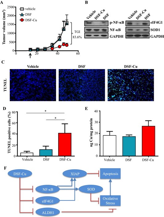
DSF‐Cu inhibits tumor growth in an in vivo model of IBC. A, Tumor volumes (measured V = (L × W2)/2) of mice with SUM149 subcutaneous flank tumors treated with vehicle, DSF, or DSF‐Cu. B, Representative immunoblot analysis of indicated proteins in tumor lysates from mice treated with vehicle, DSF, or DSF‐Cu. GAPDH as loading control. C, Representative images of tumor tissue from mice treated with vehicle, DSF, or DSF‐Cu with TUNEL staining. Magnification: 40×. DAPI (nuclei): blue; TUNEL (apoptotic cells): green. D, Quantification of TUNEL positive cells (from C). Mean ± SEM % TUNEL positive/total number of cells, *p < 0.05. E, Cu content of excised tumors (ng) measured by ICP‐HRMS relative to protein (mg). F, Schematic representation of DSF‐Cu mechanisms of action. DSF‐Cu complex acts as a pro‐oxidant, induces ROS‐mediated cancer cell death by inhibiting NFκB, which attenuates NFκB‐dependent antioxidant and anti‐apoptotic gene expression. DSF‐Cu inhibits ALDH1, which has been implicated in protection from ROS. DSF‐Cu also inhibits the potent anti‐apoptotic protein, XIAP, and translation initiation factor eIF4G1 (which can enhance XIAP translation during cell stress), promoting apoptosis.