
Figure 1. Aging and lack of Nrf2 increases oxidative stress in skeletal muscle.
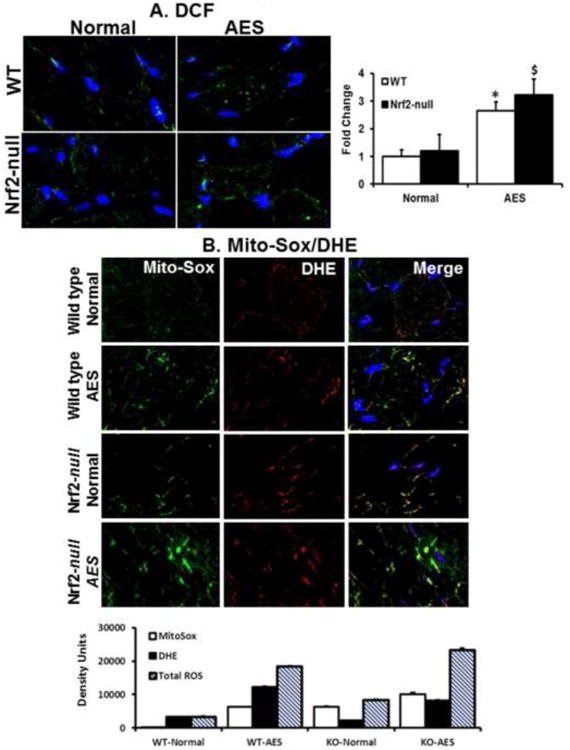
A. Determination of ROS in the skeletal muscle tissue sections using fluorescent (dihydro difluro diacetate (H2DCFDA)/dihydroethidium (DHE) probes and microscopy. Frozen tissue sections were incubated with 10μM of H2DCFDA for 30 min at 37° C. Sections were then washed with 1XPBS, mounted and analyzed by Zeiss 510 Meta confocal microscopy. Significantly increased ROS levels were seen in the WT and Nrf2-null mice in response to AEES compared with SED at 23 months (p<0.01), but these values were comparable between WT and Nrf2-null under sedentary condition.
B. Determination of the source of ROS generation using mitosox green and DHE fluorescent probes. Frozen sections of SM were stained with 10μM mitosox green and dihydroethidium (DHE) for 30 minutes and imaged using confocal microscope. Images from green and red fluorescence were merged (purple) to locate the mitochondrial source of ROS generation. The purple color is indicating ROS within the mitochondria. Both AEES and abrogation of Nrf2 showing increased ROS generation in the mitochondria of SM. Histograms showing density of the fluorescent signals from WT and Nrf2-null mice under sedentary and AEES conditions. The data are from n=3 mice/group and *p<0.05.