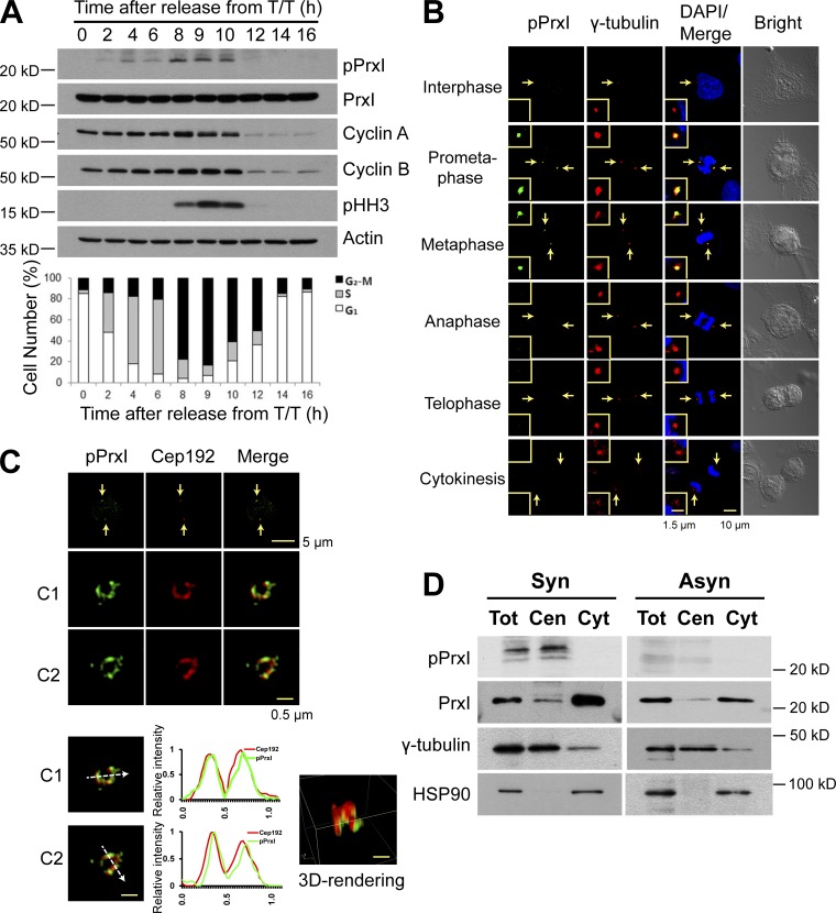
Figure 1.
Phosphorylation of PrxI at Thr90 occurs at the centrosome of HeLa cells during early mitosis. (A, top) HeLa cells that had been arrested at the G1–S border with a double thymidine block (T/T) were released in fresh medium (at 0 h) and collected at the indicated times for immunoblot analysis with antibodies to the indicated proteins. (bottom) The percentage of cells in the various phases of the cell cycle was estimated by flow cytometric analysis. Data are representative of three experiments with similar results. (B) Confocal microscopy of asynchronously growing HeLa cells stained with antibodies to pPrxI (green) and to γ-tubulin (red). Cell cycle stage was monitored by staining of DNA with DAPI (blue) and bright-field imaging. The areas indicated by the arrows are shown at higher magnification in the insets. (C) 3D-SIM images of pPrxI (green) on two pericentrosomes (C1 and C2) of mitotic HeLa cells. Fluorescent line intensity histograms show the colocalization of pPrxI (green) with pericentrosomal protein Cep192 (red) in two centrosomes. 3D-rendering image is shown in one centrosome. The data shown are from a single representative experiment out of three repeats. Arrows indicate pericentrosomes. (D) HeLa cells synchronized at prometaphase by treatment with thymidine and nocodazole (synchronous [Syn]) or those growing asynchronously (Asyn) were subjected to subcellular fractionation. Total cell lysates without nuclei (Tot) as well as cytosolic (Cyt) and centrosome (Cen) fractions were subjected to immunoblot analysis with antibodies to the indicated proteins.

