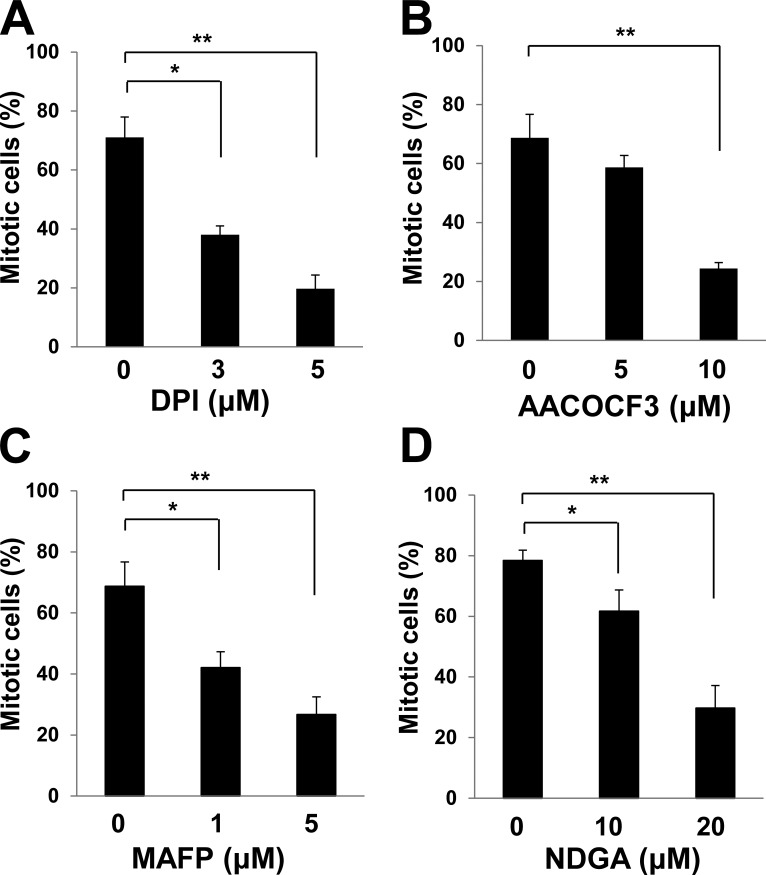
Figure 3.
Sources of H2O2 produced during mitotic entry. (A–D) HeLa cells stably expressing histone H2B–GFP were synchronized at G1–S by thymidine treatment for 18 h, released into S phase in thymidine-free medium for 3 h, and then incubated in medium containing nocodazole and various concentrations of DPI (A), AACOCF3 (B), MAFP (C), or NDGA (D) for 10 h. The percentage of mitotic cells was then estimated on the basis of chromosome condensation and cell rounding. Data are means ± SD from three independent experiments. *, P < 0.05; **, P < 0.005 (Student’s t test).

