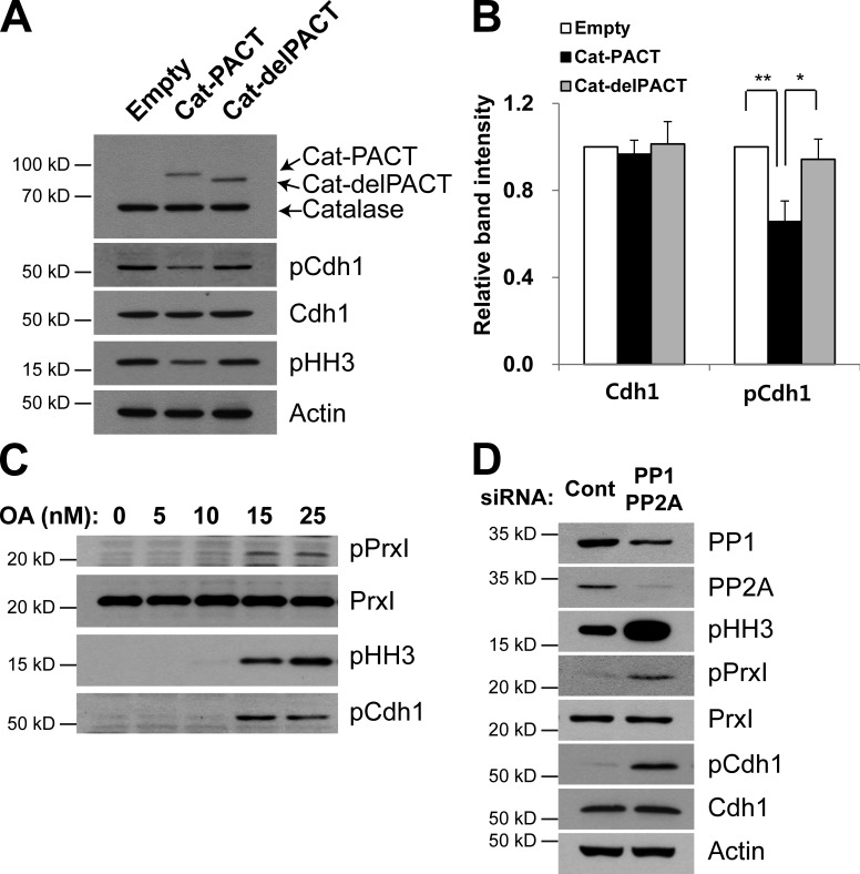
Figure 4.
Effects of centrosome-targeted catalase and OA on Cdh1 phosphorylation and mitotic entry. (A and B) HeLa cells were infected with empty, Cat-PACT, or Cat-delPACT retroviral vectors and synchronized at prometaphase. Cell lysates were then subjected to immunoblot analysis with antibodies to the indicated proteins (A). The relative immunoblot intensities of Cdh1 and pCdh1 normalized by those of actin were determined as means ± SD from three independent experiments (B). *, P < 0.05; **, P < 0.005 (Student’s t test). (C and D) HeLa cells were cultured in the presence of the indicated concentrations of okadaic acid (OA) for 24 h (C) or transiently transfected for 48 h with control siRNA or with a mixtures of siRNAs for PP1, PP2Aα, and PP2Aβ (D), after which cell lysates were subjected to immunoblot analysis with antibodies to the indicated proteins. Data are representative of three experiments with similar results. Cont, control.

