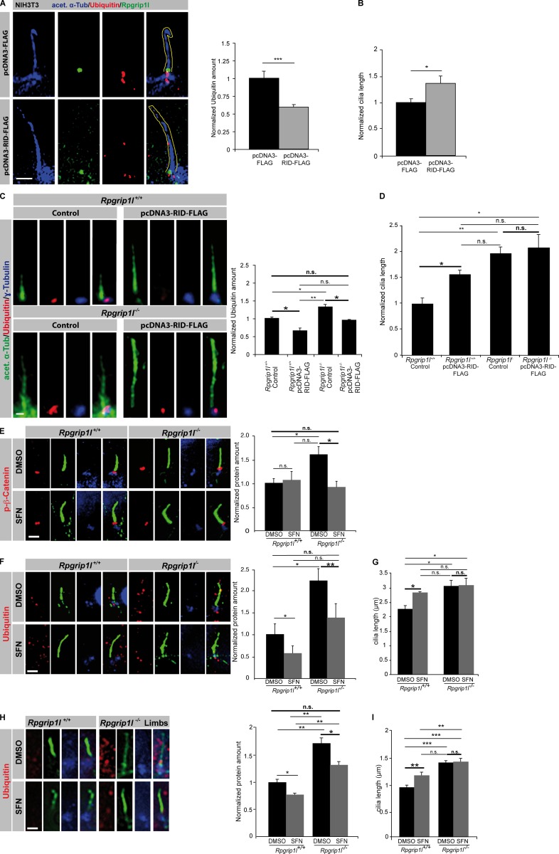
Figure 10.
Transfection of the RID domain and SFN treatment increase proteasomal activity and rescue the decreased activity of the ciliary proteasome caused by Rpgrip1l deficiency. (A and B) Immunofluorescence on NIH3T3 cells. Per control and per transfected cells, 20 cilia were used for quantification. The ciliary axoneme is marked by acetylated α-tubulin (acet. α-Tub). (A) Yellow lines encircle the shape of cilia. (C–I) The most important significance comparisons are written in bold with larger asterisks as are larger not significant comparisons. (C and D) Immunofluorescence on MEFs of E12.5 WT and Rpgrip1l−/− embryos (both genotypes, n = 3 embryos). At least 10 cilia per embryo were used for quantification. The ciliary axoneme is marked by acetylated α-tubulin (acet. α-Tub) and the BB by γ-tubulin. (E–G) Immunofluorescence on MEFs isolated from E12.5 WT and Rpgrip1l−/− embryos (both genotypes: p-β-Catenin and Ubiquitin [treated with DMSO or SFN], n = 3 embryos, respectively). Per embryo, 20 cilia were used in the quantifications. The ciliary axoneme is marked by acetylated α-tubulin (green), and the BB is marked by γ-tubulin (blue). All quantified proteins are shown in red. (H and I) Whole embryo culture treatment of embryos isolated on E12.5 and incubated for 5 h in 20 µmol SFN (both genotypes: Ubiquitin [treated with DMSO or SFN], n = 3 embryos, respectively]). Per embryo, 20 cilia were used in the quantifications. The ciliary axoneme is marked by acetylated α-tubulin (green), and the BB is given by γ-tubulin (blue). Bars, 1 µm. Error bars show standard error of the mean. *, P < 0.05; **, P < 0.01; ***, P < 0.001.

