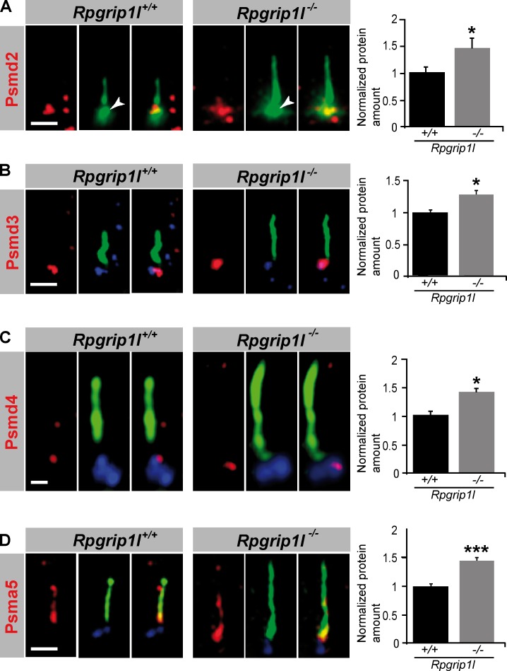
Figure 6.
Rpgrip1l deficiency results in an accumulation of proteasomal subunit components at the base of cilia. (A–D) Immunofluorescence on MEFs of E12.5 WT and Rpgrip1l−/− embryos (both genotypes: Psmd2, n = 7 embryos; Psmd3, n = 3 embryos; Psmd4, n = 3 embryos; Psma5, n = 5 embryos). At least 10 cilia per embryo were used for Psmd2, Psmd3, and Psmd4 quantification, respectively, and 20 cilia per embryo were used for Psma5 quantification. The ciliary axoneme is marked by acetylated α-tubulin (green). The BB is marked by Pcnt (green; white arrowheads; A) or γ-tubulin (blue; B–D). Error bars show standard error of the mean. *, P < 0.05; ***, P < 0.001. Bars: (A, B, and D) 1 µm; (C) 0.5 µm.

