Figure 8.
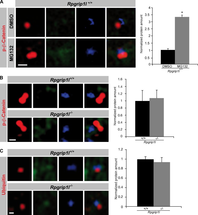
Proteasomal activity is unaltered at Rpgrip1l−/− centrosomes. (A–C) Immunofluorescence on MEFs isolated from E12.5 WT and Rpgrip1l−/− embryos (WT: p-β-Catenin (treated with DMSO or MG132): n = 3 embryos; both genotypes: p-β-Catenin and Ubiquitin: n = 3 embryos, respectively). The ciliary axoneme is marked by acetylated α-tubulin (green) and the centrosomes (basal bodies in case of ciliary presence) by γ-tubulin (blue). All quantified proteins are shown in red. An axonemal-like green staining is not visible, demonstrating that the blue staining marks centrosomes. (A) After treatment of WT MEFs with the proteasome inhibitor MG132, the amount of phospho-(S33/37/T41)-β-Catenin is significantly increased at the centrosome. (B and C) The amounts of phospho-(S33/37/T41)-β-Catenin and Ubiquitin are unaltered at the centrosome of Rpgrip1l−/− MEFs. (A–C) Per embryo, 20 cilia were used in the quantifications. Error bars show standard error of the mean. *, P < 0.05. Bars, 0.5 µm.

