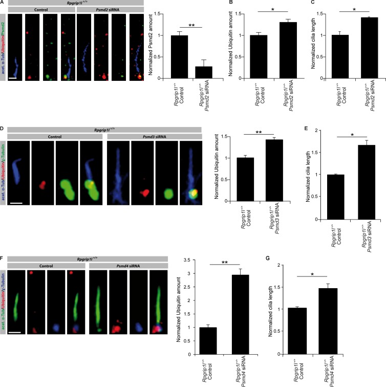
Figure 9.
Knockdown of the 19S proteasomal subunit components Psmd2, Psmd3, and Psmd4 reduce the activity of the ciliary proteasome. (A–G) Immunofluorescence on MEFs of E12.5 WT embryos (both genotypes: n = 3 embryos). (A) The ciliary axoneme is marked by acetylated α-tubulin (acet. α-Tub). Transfection of MEFs with siRNA against Psmd2 and quantification of the ciliary Psmd2 amount (A) and the ciliary Ubiquitin amount (B). (C) Measurement of cilia length after treatment with siRNA against Psmd2. (D and E) Transfection of MEFs with siRNA against Psmd3. (D) Quantification of Ubiquitin amount at cilia. The ciliary axoneme is marked by acetylated α-tubulin (acet. α-Tub)and the BB by γ-tubulin. (E) Measurement of cilia length. (F and G) Transfection of MEFs with siRNA against Psmd4. (F) Quantification of Ubiquitin amount at cilia. The ciliary axoneme is marked by acetylated α-tubulin (acet. α-Tub) and the BB by γ-tubulin. (G) Measurement of cilia length. Error bars show SEM. *, P < 0.05; **, P < 0.01. Bars: (A) 0.5 µm; (D and F) 1 µm.

