Figure 3.
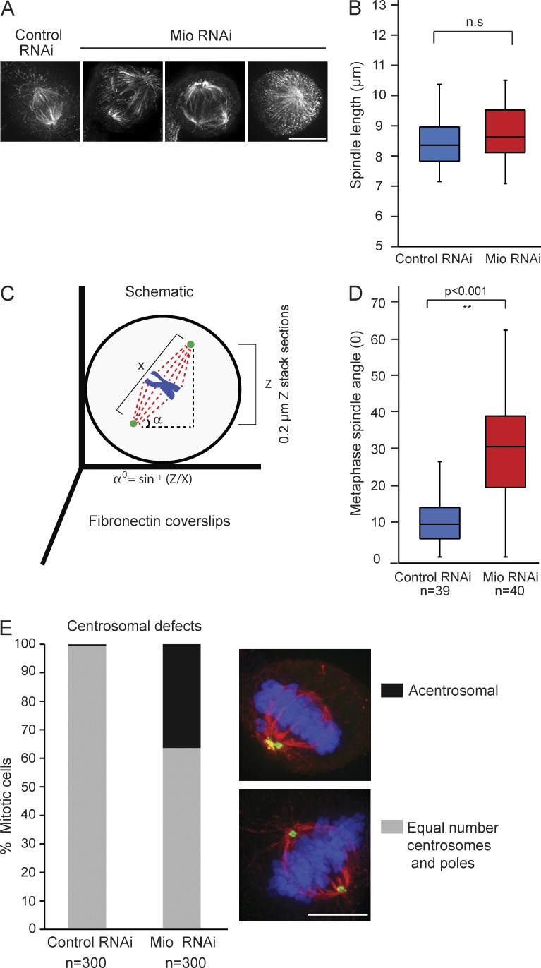
Mio depletion leads to spindle misorientation and centrosomal defects. (A) Abnormal spindle morphology detected after Mio depletion. HeLa cells were transfected with control and Mio siRNA oligos for 48 h and stained with α-tubulin. (B) Quantification of metaphase spindle length between control (blue) and Mio-depleted cells (red) shows no significant change in the length of the mitotic spindle. (C) Schematic depicting the spindle angle (α) measurement relative to the fibronectin substratum. (D) Quantification of metaphase spindle angles between control (blue) and Mio-depleted cells (red) showing a significant increase of >20o of spindle angle. n = 40 from three experiments. (E) Quantification of mitotic metaphase cells with an unequal number of centrosomes and spindle poles. 3D maximum intensity projections of representative metaphase cells immunostained with α-pericentrin (green), α-tubulin (red), and DNA (blue) are shown on the right. 300 cells per condition (n = 3). Bars, 10 µm.

