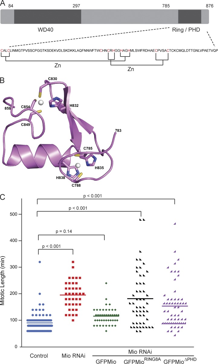
Figure 8.
Mitotic progression is impaired in the absence of the Ring/PHD domain. (A) Schematic diagram showing the domain structure of Mio and the sequence of its C-terminal Ring/PHD domain. The predicted cysteines and histidines involved in zinc coordination are labeled in red. (B) Cartoon representation of predicted 3D molecular structure of Mio C-terminal Ring/PHD domain showing the mutated residues in stick form. Zinc ions are shown in ball representation (as white spheres). (C) Mitotic progression scatter plots with NEBD as T = 0 in control (blue), Mio siRNA-treated cells with and without (red) expression of siRNA-resistant versions of wild-type GFP-Mio (green), GFP-MioRing8A (black), and GFP-MioΔPHD (purple) from live cell videos. n = 214 cells from two independent experiments. Statistical significance was determined by an unpaired Student’s t test.

