
Figure 1.
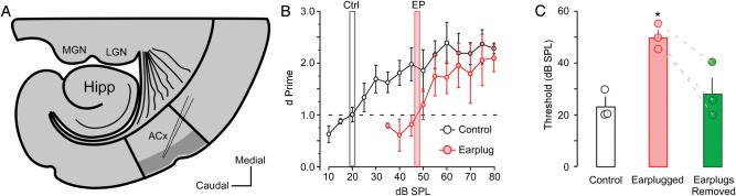
Thalamocortical slice preparation and auditory thresholds. (A) Diagrammatic sketch illustrates the thalamocortical slice preparation and L2/3 sampling region where whole-cell recordings were conducted (gray area). MGN: medial geniculate nucleus, LGN: lateral geniculate nucleus, Hipp: Hippocampus, and ACx: primary auditory cortex. (B) Line plot shows behaviorally assessed auditory thresholds for control animals (gray bar; n = 3) and animals that received earplugs on P11 (red bar; n = 3). Dashed line represents a threshold criterion of d′ = 1. (C) Bar graph shows the group average and individual auditory threshold for control animals, and earplug animals before and after earplug removal. * = P < 0.05.