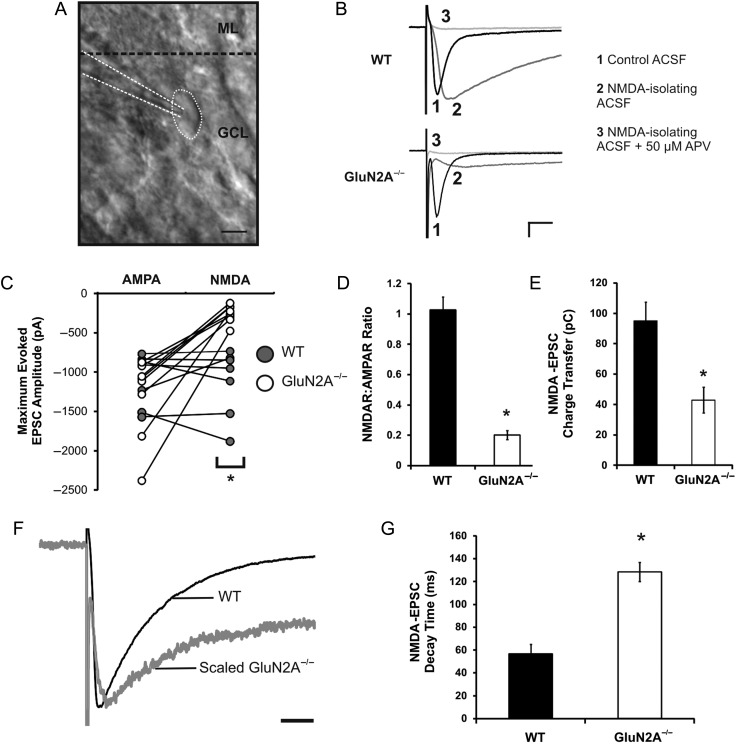
Figure 1.
Reduced NMDA:AMPA ratio and increased NMDA receptor decay rate in GluN2A−/− mice. (A) Image of a dentate granule cell, taken at ×40 magnification using oblique illumination (ML, molecular layer; GCL, granule cell layer). (B) Representative traces of EPSCs from WT and GluN2A−/− synapses, recorded in control ACSF (1), low-Mg2+ (0.1 mM) ACSF with NBQX (5 μM) and glycine (20 µM) (NMDA-isolating ACSF) (2), and low-Mg2+ ACSF with NBQX, glycine, and APV (50 μM) (NMDA-isolating ACSF + APV) (3). (C) Maximum amplitudes of AMPA and NMDA-EPSCs in WT and GluN2A−/− cells revealed a significant reduction in NMDA-EPSCs at GluN2A−/− synapses. (D) The NMDA:AMPA EPSC ratio was severely reduced at GluN2A−/− synapses. (E) Charge transfer via NMDA receptors was significantly reduced at GluN2A−/− synapses. (F) Representative traces of a GluN2A−/− NMDA-EPSC (gray), scaled to a WT NMDA-EPSC (black). (G) Weighted decay time constant of NMDA-EPSCs was significantly increased at GluN2A−/− synapses (constant generated through double exponential fit of decay phase). In all graphs, wild-type (WT): black; GluN2A−/−: white. Data are represented as mean ± SEM. *P < 0.05 denotes statistical difference. Scale bar for A = 10 μm, scale bar for B = 200 pA, 20 ms, scale bar for F = 50 ms.

