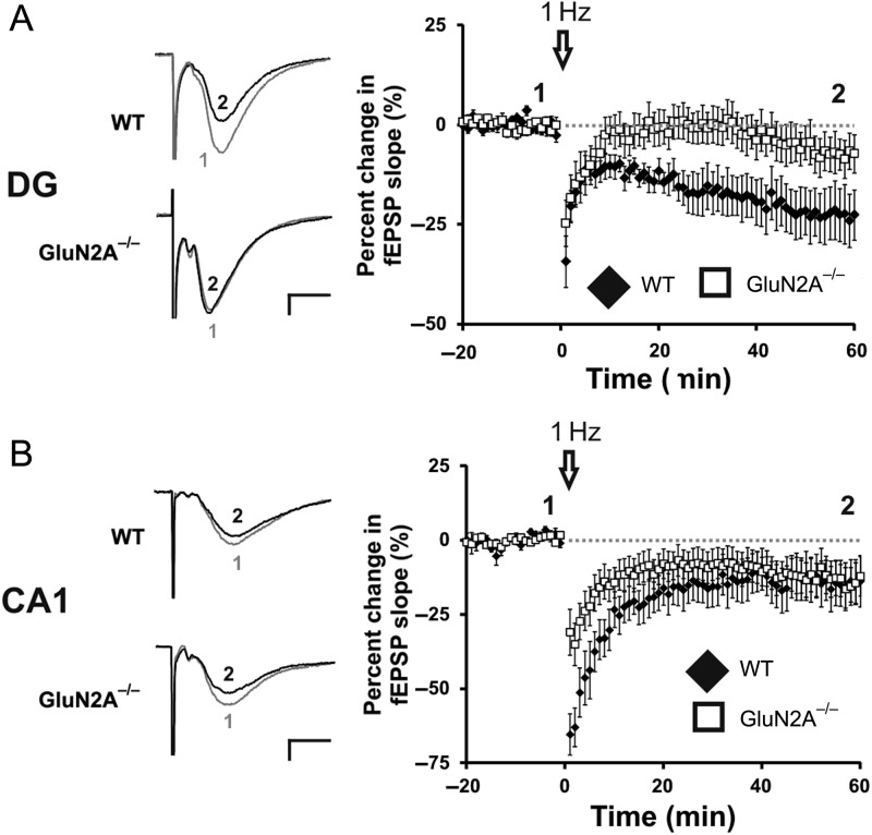
Figure 4.
GluN2A−/− mice have abolished LTD in the DG, intact LTD in the CA1. (A) Lack of LTD induced through a LFS conditioning protocol (1 Hz, 900 pulses) at GluN2A−/− synapses in the DG. Representative traces from WT and GluN2A−/− DG slices, before (1) and after (2) the low-frequency conditioning protocol. (B) Normal LTD induced through a low-frequency conditioning protocol (1 Hz, 900 pulses) at GluN2A−/− synapses in the CA1. Representative traces from WT and GluN2A−/− DG slices, before (1) and after (2) the low-frequency conditioning protocol. In all graphs, wild-type (WT): black; GluN2A−/−: white. Data are represented as means ± SEM. *P < 0.05 denotes statistical difference. Scale bar = 0.2 mV, 5 ms.

