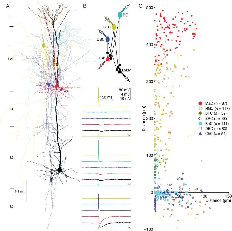
Figure 5.
L2/3 interneurons target different compartments of L2/3 pyramidal neurons. (A) Reconstruction of L2 BTC (yellow), L2 BaC (cyan), L3 DBC (blue), and L3 and L5 pyramidal neurons (red and black) recorded simultaneously from an acute cortical slice. The double colored dots indicate the putative synaptic contacts based on anatomical reconstruction. (B) Single action potentials elicited in presynaptic BTC, BaC and DBC evoked uIPSPs in postsynaptic L3 and L5 pyramidal neurons, respectively. The above schematic drawing shows symbolically the synaptic connections. Scale bars apply to all recording traces with 80 and 4 mV bars applied to traces with and without action potentials, respectively. (C) The coordinates, or the horizontal and vertical distance of the synapses made by seven groups of L2/3 interneurons from the soma of L2/3 pyramidal neurons (see Table 19 for values).

