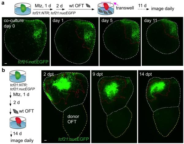
Extended Data Figure 6. Context-specific effects of outflow tract on epicardial regeneration.
a, (Top) Following ex vivo epicardial ablation and BA removal, ventricles were co-cultured with 10 outflow tracts in a transwell assay and observed for regeneration. (Bottom) No evidence for epicardial regeneration was observed in these experiments (n = 9; behavior seen in all samples). b, (Left) Following ex vivo epicardial ablation and BA removal, a non-transgenic BA (labeled as donor OFT) was transplanted to the apex and observed for regeneration. (Right) No evidence for regeneration of EGFP+ epicardium from apex to base was observed in these experiments (n = 10; behavior seen in all samples). Red dashed lines in (a, b), epicardium. White dashed lines in (a, b), ventricle. Yellow dashed lines in (b), donor outflow tract. Scale bars, 50 μm.

